What is bright field, phase contrast, dark field, DIC microscope
Time: 2018-05-19 Reads: 396833 Edit: Admin
What is bright field, phase contrast, dark field, polarizing, DIC microscope
Bright field microscope is the simplest of all the optical microscope illumination techniques. Sample illumination is transmitted (i.e., illuminated from below and observed from above) white light and contrast in the sample is caused by absorbance of some of the transmitted light in dense areas of the sample.
Phase-contrast microscope is an optical-microscope technique that converts phase shifts in light passing through a transparent specimen to brightness changes in the image. Phase shifts themselves are invisible, but become visible when shown as brightness variations.
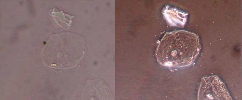
The following picture is cell under biological microscope, shows the different of phase-contrast microscope and bright field microscope. The left one is by bright field, the right one is by phase-contrast.
Phase-contrast microscope is an optical-microscope technique that converts phase shifts in light passing through a transparent specimen to brightness changes in the image. Phase shifts themselves are invisible, but become visible when shown as brightness variations.
The following picture is cell under biological microscope, shows the different of phase-contrast microscope and bright field microscope. The left one is by bright field, the right one is by phase-contrast.
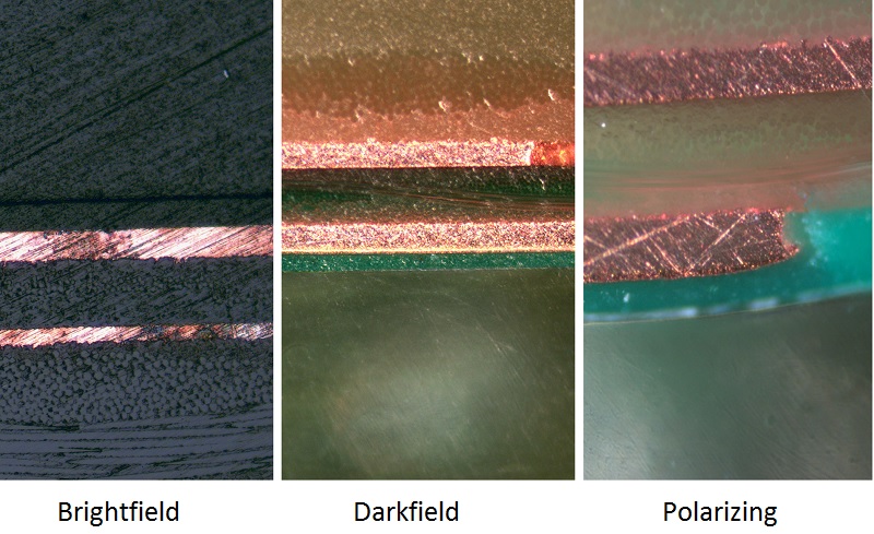
Dark-field microscope (dark-ground microscope) describes microscope methods, the field around the specimen (i.e., where there is no specimen to scatter the beam) is generally dark. The following picture shows the different of dark field, phase-contrast biological microscope and bright field biological microscope.
.jpg)
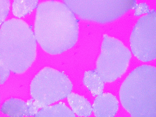
Polarizing microscope: Polarized light microscope means optical microscope techniques involving polarized light. Directly transmitted light can be blocked with a polariser orientated at 90 degrees to the illumination. The following picture is by metallurgical polaring misroscope. The sample is drug powder.
The following picture shows the difference of polarizing and darkfield metallurgical microscopes. The sample is PCB section.
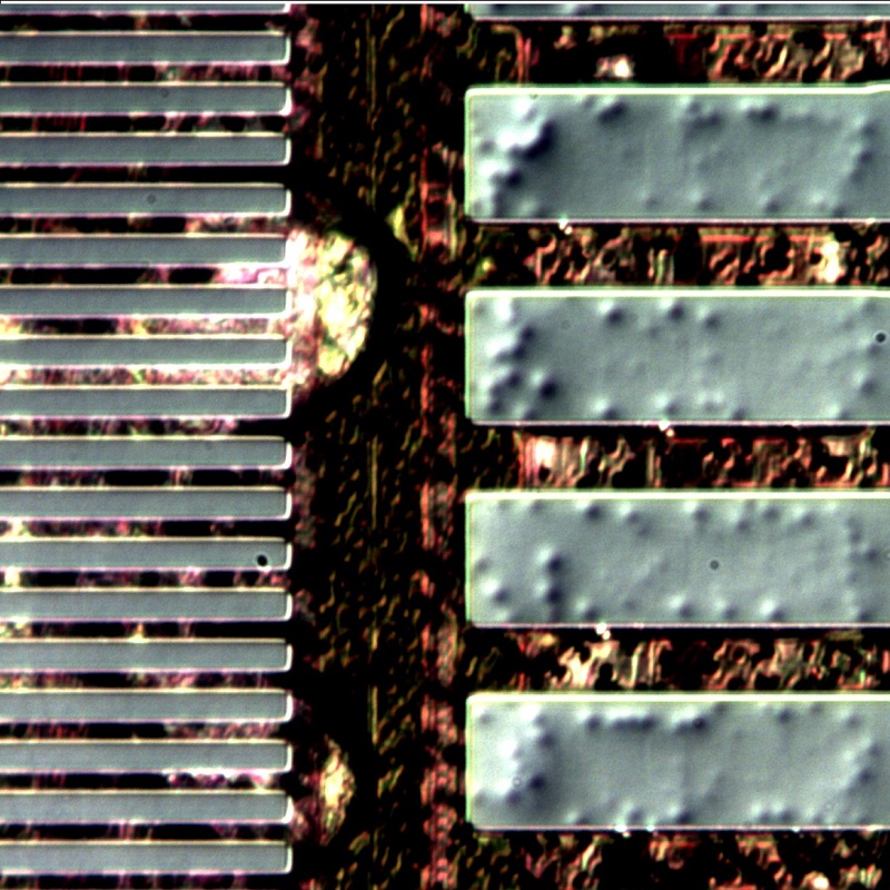
Differential interference contrast (DIC) microscope, is an optical microscope technique used to enhance the contrast in unstained, transparent samples. DIC works on the principle of interferometry to gain information about the optical path length of the sample, to see otherwise invisible features. The following picture is by metallurgical DIC microscope.